
What Causes Red Moles on Skin and How to Get Rid of Them
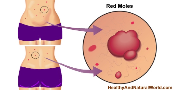
Finding a red mole on your body can be a cause for alarm but mostly it’s nothing to worry about. These red skin growths can occur anywhere on the skin and can have the appearance of bright cherry-red bumps. The red color in these moles usually comes from the cluster of blood vessels that build up near the skin’s surface. The most common type of red moles is cherry angiomas which are sometimes called Campbell De Morgan spots or senile angiomas. They usually grow on the trunk area and increase in number with age.
The good news is that red moles are usually harmless growths; however, you should always have any kind of new growth on your skin checked out by a doctor.
In this article, you will find out about the most common types of red moles and what you can do about them. In general, doctors don’t recommend to try and remove red moles by yourself as they need to be checked by a dermatologist first. Dermatologists use a variety of methods to get rid of red moles including freezing them, removing them using laser, or cauterizing them. Many people have successfully removed harmless moles at home using ingredients such as apple cider vinegar (ACV) or food grade hydrogen peroxide and you will find out how to do it towards the end of the article.
What Are Red Moles?
Most types of red moles are classed as benign skin growths with the most common being cherry angiomas. According to the American Osteopathic College of Dermatology, red moles are formed by dilated blood vessels that form into a growth.1
Red moles can be of various shapes and sizes. Dr. David Swanson on MedlinePlus says that some red moles can range from the size of a pinhead to about one-quarter of an inch. Depending on the number of blood vessels involved, the head can be smooth or stick out from the skin.2
Some other types of red moles are called actinic keratosis which are caused by exposure to the sun’s harmful UV rays and should be monitored more closely.
Let’s look a bit more closely at the various kinds of red skin growths that can appear on your body. We will then look at when red moles need to be removed and how to remove them.
The Different Types of Red Moles and What Causes Them
Different types of red moles, red skin tags, and skin growths affect many people. Here are the main types of red moles that can grow on your body.
Cherry angiomas
Cherry angioma (also known as Campbell De Morgan spot or senile angioma) is a cluster of blood vessels just under the skin’s surface that can cause red moles. As their name suggests, cherry angiomas are bright red like a ripe cherry.
According to the Primary Care Dermatology Society, cherry angiomas or Campbell De Morgan spots commonly affect the upper torso and are more common in people over the age of 40. Depending on the way the blood vessels cluster together, the moles can also be blue or purple, not just bright red.3
It’s not just your trunk area that cherry-red angiomas can affect. The journal Case Reports in Dermatology says that cherry angiomas also cause red papules to occur on the scalp. These red skin spots can also itch and may bleed if scratching them damages the skin’s surface.4
It is not known what causes senile angiomas or cherry angiomas. However, doctors know that there are some factors that can increase the number of cherry-colored skin lesions. For example, it is known that red moles occur in middle-aged people more often than in younger people.
Doctors from the American Osteopathic College of Dermatology say that pregnancy can increase the number of red-colored angiomas on the upper body. However, if you have many red bumps on your skin, it could be one of the signs of liver damage.1
Some researchers have found a link between exposure to bromides and the appearance of cherry angiomas. The journal Dermatology reported that two lab technicians who were exposed to compounds containing bromide developed a number of cherry angiomas on their trunks.5 Bromide has been used in some medications and for scientific purposes.6
Actinic keratosis
Some types of reddish-colored moles can be caused by too much exposure to the sun. Actinic keratosis is characterized by red skin growths that have a wart-like surface on the nodule.
Doctors from the Mayo Clinic say that some of the other symptoms of actinic keratosis are itching or burning around the skin growth, the growth has a hard surface, and the skin lesion is a red, pink, or brown color. Actinic keratosis can become cancerous and it’s important to have any red, hard skin growths checked out by a doctor.7
Spitz nevus
Spitz nevus is another type of red mole that looks like a dome-shaped red bump on the skin. The most common places for Spitz nevi red nodules to grow are on the limbs and face.
Dermatologists from DermaNet New Zealand say that Spitz nevi are similar in appearance to melanoma. However, unlike melanoma growths, Spitz nevi are benign. Because they are similar to cancerous skin growths, you should always have a qualified dermatologist check out the red moles.8
Apart from affecting the upper trunk, the American Osteopathic College of Dermatology says that red Spitz nevi moles can also affect the neck, and arms. The red moles can become itchy and bleed if the surface of the skin bump is broken.9
How to Prevent Red Moles
Red moles like cherry angiomas and Spitz nevi are not always preventable. As I already mentioned, cherry angiomas are mainly connected with getting older and some pregnant women also develop them. However, according to the American Osteopathic College of Dermatology, a large number of red moles in the form of cherry angiomas or spider angiomas are connected with liver damage in some people.1
Your liver plays an important function in your body and it’s important to keep your liver in good health. This helps to keep your blood healthy and free of toxins.
It’s important to prevent red moles and skin growths that are connected with sun damage. Actinic keratosis can lead to skin cancer and you should always keep your skin protected in strong sunshine. If a red mole that’s associated with actinic keratosis starts to grow or bleed, you should have a doctor look at it as soon as possible.
Natural Ways to Remove Red Moles
Should you remove red moles at home?
Dermatologists don’t recommend removing red moles at home unless they have been examined and assessed by a professional.
However, if a dermatologist has determined that the red mole is benign, there are a couple of natural ways that you can try remove them.
Apple cider vinegar (ACV)
Apple cider vinegar is a natural remedy to get rid of unwanted skin growths such as red moles. Apple cider vinegar is naturally acidic and it can help to gently get rid of skin tags, warts, and possibly cherry angiomas. If you want to use apple cider vinegar for its medicinal purposes, you should always use raw, unprocessed apple cider vinegar.
Don’t use ACV to remove red moles around the eyelids, as it can harm the eyes.
How to use:
To use apple cider vinegar for red mole removal, this is what you should do:
- Dip a small piece cotton ball in raw apple cider vinegar.
- Place the apple cider vinegar soaked cotton ball on the red mole and secure with a band aid. Make sure that the cotton ball only covers the red mole to prevent any irritation to the skin around the mole.
- Leave on overnight and remove it in the morning.
- Continue applying the ACV remedy to the cherry angioma every night for around a week until it falls off.
For more information, please read my article on how to use apple cider vinegar (ACV) to remove skin tags and warts.
Hydrogen peroxide
Hydrogen peroxide is another natural remedy that may be helpful in removing cherry angiomas. Hydrogen peroxide can be used to help get rid of skin tags and warts naturally because it is a natural bleaching agent. You should use 35% food-grade hydrogen peroxide for wart and skin growth removal.
How to use:
If a dermatologist has given you the “all clear” on your cherry angiomas, you can use 35% hydrogen peroxide to remove the red mole this way:
- Using a file, gently rub away the top layer of skin over the red mole.
- Soak the end of a cotton swab with food-grade hydrogen peroxide.
- Carefully rub the solution onto the red mole making sure that you don’t get it onto surrounding skin.
- Repeat 3 to 5 times a day for around 2 weeks to get rid of the red cherry angioma.
Please read my article on using hydrogen peroxide to remove skin tags with hydrogen peroxide safely to find out more information.
Medical Options to Get Rid of Red Moles
In many cases, red moles such as cherry angiomas are benign and don’t need any treatment. However, for cosmetic reasons many people choose to get rid of these red moles. However, a dermatologist will recommend the best kind of treatment.
Some of the conventional ways that dermatologists remove red moles are as follows:
Cryosurgery. Cryosurgery involves freezing off the red mole with liquid nitrogen. The Journal of Dermatologic Surgery and Oncology says that Cryosurgery is often used to remove cherry angiomas for cosmetic reasons. There can be some discomfort associated with this; however, dermatologists will administer local anesthetic.10
Electrocauterization. Electrocauterization is a process of burning of unwanted red skin growths that is also used to get rid of cherry angiomas. Very often lasers are used to precisely cut off the red papule. The Journal of the American Academy of Dermatology reported that laser cauterization can successfully remove red moles. Sometimes, many people experience inflammation around the treated area.11
Pulsed dye laser: Similar to electrocauterization, laser is used to burn off the red mole. It does it by concentrating the heat of a focused laser beam on the skin growth.
Curettage. Curettage is a way to scrape off unwanted growths and cancerous moles. A dermatology journal says that curettage is an acceptable way to treat cherry angiomas.4
Sclerotherapy. A cheap way that some dermatologists use to remove red moles is by injecting a saline solution. The journal Dermatological Surgery reported that sclerotherapy is a useful treatment for cherry angiomas. The majority of cases were cured with 3 weeks of regular injections.12
How to Tell if a Red Mole is Dangerous
In general, doctors recommend keeping an eye on any moles to observe if they change in any way. However, if you notice a new skin growth that is red, brown, black, or pinkish, you should have it checked out by a dermatologist. To check for moles that are changing and could become cancerous, you should follow the ACBDEs. This is:
- Asymmetry. The mole has an irregular shape and one half isn’t the same shape and size as the other half.
- Border. Worrisome moles will have a ragged or irregular border.
- Color. The mole has different shades of brown, black, white, blue, or white.
- Diameter. The size of the mole is larger than a pencil’s eraser.
- Evolves. The mole changes in shape, size, and color.
If a mole exhibits any of these traits, it requires a close look by a physician to be safe. New moles that grow in quickly and any that itch and bleed should also be taken into consideration.
Other warning signs of skin cancer that you should look for are:
Persistent Sores – When inflammatory sores form and refuse to go away despite treatment attempts.
Bruises and Aching – Melanoma may also come in the form of bruised and off colored skin that lingers much longer than a normal bruise. Some areas of the skin might be tender and randomly ache throughout the day.
Check Your Fingernails – Sometimes melanoma can signal its development with the formation of black and brown marks underneath the toe and fingernails. Read also the article about 11 health warnings your fingernails may be sending.
For more information, please read the article: Warning Signs of Melanoma, The Most Deadly Form Of Skin Cancer.
Read these related articles:
Article Sources